Facebook
 Google
 GitHub
 Linkedin
Ultrasound Receiver Architecture Undergoes a Seismic Shift
Ultrasound, with nearly a century of technological history, has seen its third major technological breakthrough coming out of North Carolina State.
Sonography, commonly called ultrasound, evolved from SONAR and entered into medical use in the 1960s, according to researchers at the Harvard Business School.
Sonography quickly gained market dominance over X-ray in the 1970s as the preferred method for diagnosing conditions in cardiology, obstetrics, and gynecology. Processing sonography data on a computer became widespread in 1985.
In the 21st-century, electronic processing systems have steadily enhanced sonography technology, improving signal-to-noise characteristics, penetration depth, gains in lateral resolution, and contrast.
Regardless of the incremental improvements from solid-state electronics, there has not been a significant shift in the basic structure of an ultrasound receiver until this year.
Researchers at North Carolina State University have announced a revolutionary new receiver for ultrasound technology that has the potential to drive down the design cost of such systems and usher in a market rush of this equipment not seen since the 1980s.

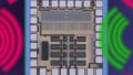
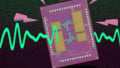
Basic diagram of the new ultrasonic imaging device. Image used courtesy of ACS Applied Materials & Interfaces
Researchers call this technology a "novel ultrasonic imaging device" because it optically displays acoustic signals on the surface of a piezoelectric transducer.
A Look at Traditional Electronic Ultrasound Systems
Ultrasound technology uses an artificially-generated sound wave in the range of 2–18 MHz, which operates on the same principles as SONAR and RADAR, where reflected waves are returned to a receiver for signal processing.
A piezoelectric effect is generated by a transducer material—in sonography, that material is lead zirconate titanate (PZT)—to transform sound waves to electrical (or electrical to sound) impulses that can be processed and displayed.

Block diagram of a traditional ultrasound electronics system showing the complex transceiver path to the transducers. Image used courtesy of Maxim Integrated
The transceiver electronics driving the sonography probe are complex, with a high-voltage transmission path (>200 Vpp) consisting of a high-voltage digital-to-analog converter, an amplifier, a transceiver switch, and a high-voltage multiplexer.
The receiver side of this system includes the following:
- Low-noise amplifier (LNA)
- Variable-gain amplifier (VGA)
- Anti-aliasing filter (AAF) used to remove components beyond the range of the first Nyquist zone of the ultrasound frequency
- Analog-to-digital converter, typically 12-bit resolution running at 60 mega-samples per second
Redefining Ultrasound Systems with Direct Processing to an OLED Display
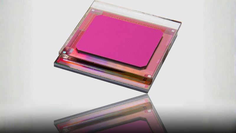
The new technology developed at NC State University seeks to remove the receiver electronics requirement in the system integration and instead replace it with a directly-processed OLED screen up to 500 px by 500 px in resolution, according to researcher Franky So.
The technology integrates a PZT material with the OLED display, which illuminates in the presence of received ultrasonic energy. “Conventional ultrasound devices have a receiver that detects ultrasonic waves and converts them into an electrical signal,” says Xiaoning Jiang. “We’ve created a device that effectively eliminates the electrical signal processing altogether.”
In the NC State research papers’ supporting information, the integrated receiver was tested with two setups: a nine-stack composite including ZnOx and an eight-stack composite without ZnOx. The variance in structure shows an improved OLED luminance and current efficiency as a function of luminance without the ZnOx.
_and_without_(right).jpg)
OLED displays with a Zinc Oxide layer (left) and without (right) showing the increased luminance between the two experimental stackups. Images (modified) used courtesy of ACS Applied Materials & Interfaces
The research team demonstrates the effects of different acoustic mediums (water, gel, and metal) on the PZT receiver as well as the effects of an electrically-opaque obstacle interfering with the receiver. The supplementary research clearly shows that the OLED display tracks the ultrasonic PZT receiver energy.
A New Era of Simply-Designed, Affordable Ultrasound?
This new technology provides a fundamentally different way to receive and process ultrasound acoustic waveforms, completely eliminating the complex electronic receiver modules found in existing ultrasound systems.
Franky So explains, “We can make ultrasound receiver-displays for $100 or so." This cost significantly contrasts conventional ultrasound technology, which costs upwards of $100,000.
Related Content