Facebook
 Google
 GitHub
 Linkedin
Ultrasound Patches Provide Continuous Imaging of Internal Organs
Researchers are refining soft adhesive ultrasound patches that provide continuous imaging—sometimes for up to 48 hours.
Most wearable health monitoring devices rely on light-based methods to provide users with biometric information—but these figures do not provide a holistic picture of a person's health.

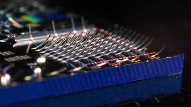
Now, wearable ultrasounds may provide continuous imaging for up to 48 hours. Image used courtesy of UCSD
One health-monitoring technology that has gained popularity in recent years is wearable ultrasound. Promising continuous monitoring and imaging of internal organs, wearable ultrasound may provide more detailed health insights than currently-available wearables.
An Ultrasound Patch Based on Doppler
In April of 2021, researchers from the Northern Ontario School of Medicine published an academic paper describing an innovation in wearable ultrasound.
The researchers presented a hands-free ultrasound patch capable of continuously monitoring the carotid artery using quantitative Doppler methods. The paper, published in Nature, describes how the Doppler patch adheres to the neck and operates using two continuous-wave 4 MHz ultrasound transducers. One transducer transmits sound continuously while the other receives echoes continuously.

Functional block diagram of the Doppler sensor. Image used courtesy of Kenny et al
The device measures frequency shifts; the frequency of the received echoes is compared to that of the transmitted beam. The change in frequency, known as the Doppler shift, is proportional to the velocity of motion toward or away from the transducer. Using this method, the patch can detect blood moving anywhere within the overlapping volume of the two beams.
The patch generates an ultrasound beam that is substantially wider than the carotid artery, allowing healthcare professionals to easily place the device over the artery with considerable tolerance. The patch adheres to the neck and allows for hands-free, consistent positioning and alignment between the ultrasound beam and the artery, creating highly accurate measurements with little manual intervention.
Measuring 14 Centimeters Deep
In July 2021, researchers from the University of California San Diego (UCSD) announced their own soft ultrasound skin patch.
Like the device developed by the Northern Ontario researchers, the UCSD patch uses ultrasound technology to monitor blood flow as well as correlated blood pressure and heart function. As described in a paper published in Nature, the hands-free patch can measure signals up to 14 centimeters deep in the body, providing extremely accurate and reliable measurements.

The ultrasound wearable wired to its full experimental setup. Image used courtesy of UCSD
The patch consists of a thin sheet of stretchable polymer that adheres to the skin and is worn on the neck or chest. The patch is integrated with an ultrasound phased array, which is a 12 x12 array of tiny ultrasound transducers, each individually controlled. The array also measures the Doppler shift of reflected ultrasound beams to visually record blood flow.
The patch has two modes of operation. In the first, the phased array transmits ultrasound waves synchronously to produce a high-intensity, focused ultrasound beam, providing a high-depth measurement. In the other mode, the array and transducers transmit out of sync, allowing ultrasound beam steering and angled measurements.
48 Hours of Continous Monitoring
The final and most recent development in wearable ultrasound comes from MIT, which announced a stamp-sized ultrasound sticker for internal body monitoring in July 2022.
The device includes an adhesive layer made of two thin layers of elastomer surrounding a middle hydrogel layer. Each layer serves a unique function: the bottom elastomer layer is designed to stick to a patient’s skin while the top layer adheres to a rigid array of transducers. Like Northern Ontario's research, the MIT device utilized Doppler shifts to detect the motion of underlying blood.

The ultrasound sticker. Image used courtesy of MIT
In their published research study, the researchers explain that the sticker stayed attached to users' skin (even on active users) and provided reliable images for up to 48 hours. The stickers were tested on multiple body parts, including the neck, chest, abdomen, and arms. Each location provided insight into the changing diameter of underlying blood vessels. Further, the stickers provided clinically-useful images of deeper organs such as the heart, stomach, and underlying muscles.
As an electrical engineer, what medtech innovations have impressed you in the past few years? Share your insights in the comments below.