Facebook
 Google
 GitHub
 Linkedin
Optical Brain Research Pushes Smaller Implants, Sharper Imaging
Cornell, MIT, and HKUST teams have reported laser- and photonics-based devices that record neurons, image deeper, and tame crosstalk in all-optical neuroscience.
Three research groups have published recent papers that lean on laser sources, photodetectors, and integrated optics to probe the brain in new ways.
Cornell engineers reported a wireless neural implant smaller than a grain of salt; an MIT-led team described a self-organizing "pencil beam" laser; and a group at the Hong Kong University of Science and Technology (HKUST) described a laser-control technique that suppresses crosstalk in all-optical neural interrogation.
Together, the projects show how optoelectronic miniaturization and beam engineering are pushing the boundaries of neuroscience tools beyond those of conventional electrode arrays and standard fiber optics.
Cornell's Salt-Grain Wireless Implant
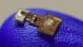
Cornell's device, called MOTE (microscale optoelectronic tetherless electrode), measures roughly 300 µm long and 70 µm wide. Power and uplink both run over light: red and infrared laser beams pass through brain tissue to energize the implant, and the device transmits recorded data back optically using pulse position modulation, the same coding used in satellite optical links.
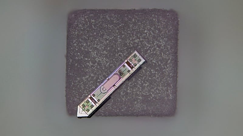
Cornell's neural implant resting on a grain of salt, measuring about 300 µm long and 70 µm wide. Image used courtesy of Cornell University
An aluminum gallium arsenide (AlGaAs) semiconductor diode captures the input light energy and emits the data signal, paired with an on-chip, low-noise amplifier and an optical encoder.
In tests reported in the paper, MOTEs implanted in the barrel cortex (the whisker sensory region) of living mice recorded action-potential spikes and broader synaptic activity for more than a year while the animals remained healthy.
Molnar said the team chose pulse-position modulation because it allows the implant to communicate with very little power while still receiving the data optically. The group is now exploring whether the device's material composition will allow recordings during MRI, with possible adaptations for the spinal cord and for opto-electronics housed in artificial skull plates.
MIT's Self-Organizing Pencil Beam
The MIT contribution didn’t start as a brain-imaging project. The research team was pushing a multimode optical fiber close to its damage threshold when the laser light, rather than becoming more chaotic with increasing power, collapsed into a single, needle-sharp beam.
The effect required two conditions: the laser had to enter the fiber at a precise 0° angle, and the drive power had to be high enough for the light to interact nonlinearly with the glass itself. Above that threshold, the nonlinearity counteracts the fiber's intrinsic disorder, yielding what the team calls a self-localized pencil beam.
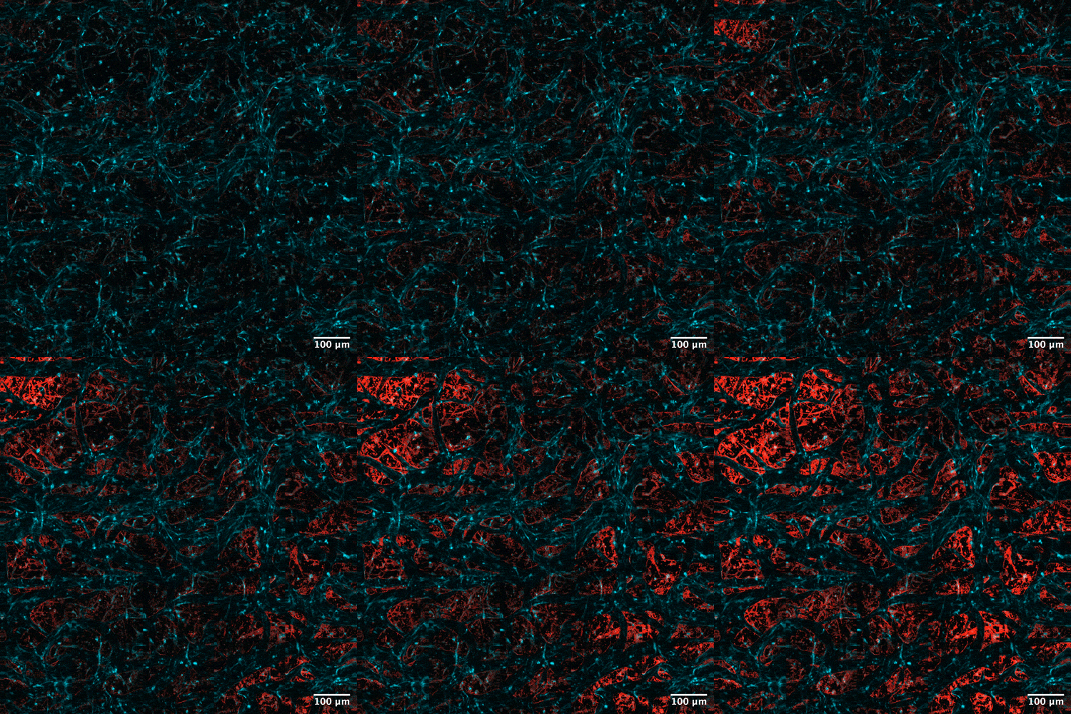
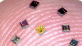
The new technique enabled researchers to track how cells absorb proteins in real-time. Drug uptake is shown in red in a blood-brain barrier model using the pencil beam. Image used courtesy of MIT
The researchers used the beam to image a human blood-brain-barrier model, capturing 3D images about 25 times faster than the gold-standard method while preserving comparable resolution and avoiding the blurry sidelobes that distort many high-power beams.
Because the technique works without fluorescent labels, the team showed individual cells absorbing drugs in real time—a capability the researchers call "a game-changer" for screening drug candidates aimed at neurodegenerative diseases such as Alzheimer's and ALS. The authors plan further work on the underlying physics and on extending the method to directly image neurons.
HKUST's Crosstalk-Free Optical Interrogation
The HKUST work addresses a different optical problem: crosstalk in "all-optical interrogation," in which genetically encoded sensors and optogenetic actuators enable researchers to both read and drive neural activity with light.
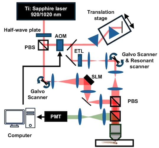
Schematic of the two-photon optical interrogation system. Image used courtesy of Nature Communications
The infrared imaging laser intended for passive recording can itself activate optogenetic proteins in nearby neurons, blurring the line between observation and stimulation. The HKUST team built a technique called Active Pixel Power Control (APPC) that acts as a per-pixel dimmer during laser scanning.
APPC uses custom mapping software to identify where optogenetic proteins are expressed and a fast acousto-optic modulator to dial down (or to zero) laser power on those specific pixels while maintaining uniform intensity elsewhere. Tested in larval zebrafish, the method preserved neuronal signal quality and suppressed optogenetic artifacts.
The HKUST team highlights that APPC is compatible with standard two-photon microscopes and can be added without replacing existing systems, and notes that the approach should extend to mouse models.